Sunday 27 March 2022

Polymerase Chain Reaction - Glossary

  1. Amplification- Increase in the number of copies of the desired DNA fragment.
  2. Annealing- Attachment of an oligonucleotide primer to the DNA or RNA template.
  3. Chromosome- Structures found in nucleus which carry the DNA molecule and the DNA holds gene.
  4. Ct (cycle threshold)- The number of cycles required for the fluorescent signal to exceed levels.
  5. Denaturation- The process of breaking of hydrogen bonds between the DNA bases in the DNA and converting DNA from double stranded to single stranded.
  6. deoxynucleotide triphosphates (dNTPs) - Building blocks of DNA synthesis which helps to grow DNA strand in the presence of Taq polymerase enzyme. N stands for A, G, T and C. 
  7. DNA - Deoxyribose Nucleic Acid is the polymer of nucleotides and it holds the genetic information of all living organisms.
  8. DNA polymerase- Enzyme which catalyzes the DNA synthesis (copies DNA into DNA) by adding nucleotides to the 3' end of the growing DNA strand. It is a DNA dependent DNA polymerase enzyme.
  9. DNA sequence - Order of nucleotides in a DNA.
  10. Enzyme- A protein which catalyzes the particular biochemical reaction without changing the nature of the reaction.
  11. Extension- The formation of new strand of DNA by Taq polymerase enzyme.
  12. Fluorescence- The process in which a compound in an excited state emits light when it returning to the ground state.
  13. Forward and Reverse Primers- Forward primer binds to the template DNA while reverse primer binds to the complementary strand.
  14. Gene- Unit of inheritance. Each section of DNA which codes for single RNA or protein.
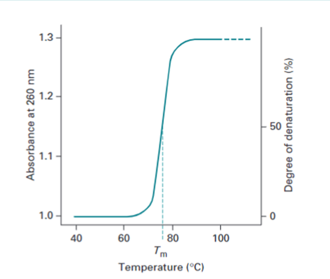
  15. Melting temperature (Tm) - The temperature at which 50% of DNA is melted (2 strands of the double stranded DNA molecule detach due to the complete breakage of hydrogen bonding).
  16. Nucleotide- A subunit of RNA or DNA consisting of nitrogenous base, a phosphate molecule and a sugar molecule.
  17. Oligonucleotide - A short sequence of nucleotide.
  18. Polymerase Chain Reaction (PCR) - PCR is a technique used in molecular biology to make many copies of the desired DNA sequence.
  19. Primer- A short oligonucleotide which is attached to the single stranded DNA molecule in order to provide a start point for strand synthesis.
  20. Replication- The process of synthesis of new copy of DNA.
  21. Reverse Transcriptase- Enzyme which catalyzes the synthesis of DNA from RNA template. It is an RNA dependent DNA polymerase enzyme.
  22. RNA- Ribonucleic acid. A single stranded nucleic acid like DNA but having uracil instead of thymine as one of the bases.
  23. Taq- A DNA polymerase commonly used in PCR, which can withstand high temperatures. 
  24. Template DNA- A precise fragment of DNA which is used as a starting material in PCR for amplification.
  25. Thermal cycler- Programmed and automated heating/cooling system for PCR applications. Enables denaturation, primer binding and extension cycles.
  26. Thermus aquaticus- Thermophilic bacterium from which Taq polymerase is purified.
References:

  • Polymerase Chain Reaction (PCR) Glossary - passel. (n.d.). Plant and Soil Sciences eLibrary. https://passel2.unl.edu/view/lesson/d81d0eedbada/glossary
  • P.Arora, M. (2005). Genetic Engineering (1st ed.). Himalaya Publishing House.
  • What does ct mean. Wisconsin Veterinary Diagnostic Laboratory. (2018). Retrieved from https://www.wvdl.wisc.edu/wp-content/uploads/2018/05/What-does-CTmeanfinahandoutlJanuary2014.pdf
            Hope you all like this information!☺ 
            Invite suggestions for more terms to elaborate the glossary!

Monday 21 March 2022

DNA Vs RNA

               Deoxyribonucleic acid and Ribonucleic acid are nucleic acids made up of nucleotides. They both are essential molecules for storing and reading genetic information in an organism.  Let us see the differences between them!

Comparison

DNA

RNA

Full form

Deoxyribonucleic acid

Ribonucleic acid

Structure

Longer chain, double-stranded arranged in double helix made up of deoxyribonucleotides

Shorter chain, single-stranded made up of ribonucleotides

Nucleotide

A nitrogenous base, 2’-deoxyribose sugar and phosphate group

A nitrogenous base, ribose sugar, and phosphate group

Function

Stores all genetic information of an organism

Involved in protein synthesis

Nitrogenous bases 

Adenine (A), Guanine (G), Thymine (T) and Cytosine (C)

Adenine (A), Guanine (G), Uracil (U) and Cytosine (C)

Nitrogenous base pairs

AT and GC

AU and GC

Location

Present in the nucleus, some amount of DNA also seen in mitochondria

Found in the nucleus, cytoplasm, and ribosome

Replication

Self-replicating

Synthesized by transcription when it is needed (DNA to RNA conversion)

Stability

More stable than RNA due to the 2’-deoxyribose. Also, thymine (5’-methyl uracil) makes DNA stable

Less stable than DNA due to the presence of the 2’-hydroxyl group which is more prone to hydrolysis

Reactivity

Less reactive than RNA, does not have any free groups to make DNA more reactive

The free 2’-hydroxyl group make RNA more reactive


References :
  • Mackenzie, R. J. (2021, November 25). DNA vs. RNA – 5 Key Differences and Comparison. Genomics Research from Technology Networks. https://www.technologynetworks.com/genomics/lists/what-are-the-key-differences-between-dna-and-rna-296719
  • Latham, K. (2021, January 22). DNA vs. RNA. Biology Dictionary. https://biologydictionary.net/dna-vs-rna/

Saturday 19 March 2022

DNA isolation- What, Why and How ?

Here, we can have a brief discussion on the fundamentals of DNA isolation.

What is DNA isolation?

The process of isolating DNA from variable sources such as blood, tissue, cell culture, buccal swab etc. A source of DNA is needed for every experiment in molecular biology, forensic, genetic labs etc. This has become a routine procedure. 

Why the DNA isolation is important?

  • To study about the cancer markers 
  • To detect genetic diseases
  • To develop new drugs
  • To detect bacterial and viral diseases
  • Paternity tests
  • Criminal investigations

How the DNA is to be isolated?

The common features of all types of DNA isolation are,


Now we can see the details of the DNA extraction,

1. Cell lysis or disruption can be done either in physical/chemical method. 
  • Physical method includes grinding/sonicating especially for plant cell (tough cell wall!). 
  • Chemical method includes treating with sodium dodecyl sulphate (SDS) or sodium lauryl sulphate (SLS) which helps in solubilizing the cell membrane.
  • Chemical method also includes the addition of enzymes which helps to digest DNA associated as well as other proteins. E.g., Proteinase K for animal cells, Lysozyme for bacterial cells etc.
2. Precipitation of DNA (separates the DNA from cell debris, detergents, proteins etc.)
  • Ice cold absolute ethanol or isopropanol is used to precipitate the DNA. DNA is insoluble in alcohol.
  • Monovalent salt such as sodium acetate is also added. The sodium ions (positively charged) react with phosphate ions in the DNA and make the DNA less hydrophilic means less soluble in water. This accelerates the precipitation process by alcohol.
  • The salt and alcohol are removed by 70% alcohol.
3. Purification of DNA
  • DNA can be re-suspended in TE buffer (Tris-EDTA buffer, pH 8).

What is Next? 

Now, we must quantify the DNA obtained. DNA quantification means checking for its purity and concentration.
  • The agarose gel electrophoresis can be used to confirm the presence of DNA.
  • UV spectrophotometer or Nanodrop can be used to determine concentration and purity of DNA. The ratio of absorbance at 260nm and 280nm (A260nm/A280nm) should be equal to 1.8 for pure DNA. If it is <1.8, protein contamination and if it is >1.8, RNA contamination.
  • By using the fact that 1 absorbance is equal to 50 µg ml-1, determine the DNA concentration using the following equation.  

50*A260nm = Concentration of DNA in Âµg ml-1

References

  • P. Arora, M. (2005). Genetic Engineering (1st ed.). Himalaya Publishing House
  • W. (2010). Principles and Techniques of Biochemistry and Molecular Biology (7th ed.). Cambridge India 
  • DNA extraction. (2009, June 18). Science Learning Hub. https://www.sciencelearn.org.nz/resources/2036-dna-extraction


Have a great day!
Thank you!

Tuesday 15 March 2022

What is DNA?

What is DNA?

DNA stands for Deoxyribonucleic acid, is the polymer of nucleotides. Every living thing has DNA, for example plants, animals, bacteria etc. Most importantly human's genetic material is also DNA, which makes us unique! DNA contains the hereditary information which passes on to you from your parents. DNA is mainly located in cell nucleus and small amount of DNA is also present in mitochondria which is known as mitochondrial DNA. 

  • DNA is long strand of 2 polynucleotides which wind one another to form a double helix structure. 
  • Each composition of a nitrogenous base, pentose sugar (known as nucleoside) and phosphate group are known as nucleotide (also known as nucleoside phosphate). Nitrogenous bases are of 4 types such as Adenine (A), Guanine (G), Cytosine (C) and Thymine (T). 
  • The bases of one strand bind with the other strand to form purine (2 ring structure) to pyrimidine (1 ring structure) that is A.T and G.C bases pairs through hydrogen bonds. 
  • Due to the bond angle of the sugar phosphate molecule, the linkages form a double helix structure.
  • Each strand of DNA has one 5'-P and 3'-OH at each end of the helix. One strand run in 5'-3' (sense strand) direction whereas other one in 3'-5' (antisense strand) which make them antiparallel.


DNA and its building blocks


Several types of bonds present in DNA double helix

  • A nucleotide or nucleoside phosphate is formed by the attachment of phosphate group to the 5' position of nucleoside by ester linkage.
  • Nucleotides are joined together by attaching 5' phosphate of one nucleotide to 3' hydroxyl of another nucleotide by phosphodiester linkage between the adjacent sugars.
  • A purine base in one strand is bonded to pyrimidine base in the another strand via hydrogen bond. Adenine pairs with Thymine by 2 hydrogen bonds and Guanine pairs with Cytosine by 3 hydrogen bonds.

Denaturation and Melting temperature

  • The loss of helical structure of DNA is known as denaturation. This occurs by the disruption of hydrogen bonds at elevated temperatures. 
  • The temperature at which half of the DNA molecules are denatured, which is known as melting temperature of DNA.

Measurement of DNA Denaturation

Melting curve of DNA

DNA denaturation is measured by using spectrophotometer. The aromatic rings in the nucleotide bases absorbs light in UV range, so the DNA also. The bases absorb 260nm light strongly.

  • The double stranded DNA has less absorbance of light at 260nm compared to single stranded DNA, because of the interference by the double helix structure of the DNA.
  • As the temperature increases DNA denatures and becomes single stranded, So absorbance increases.

References

  • Alberts B, Johnson A, Lewis J, et al. Molecular Biology of the Cell. 4th edition. New York: Garland Science; 2002. The Structure and Function of DNA. Available from: https://www.ncbi.nlm.nih.gov/books/NBK26821/
  • What is DNA? (2022, February 18). Yourgenome. https://www.yourgenome.org/facts/what-is-dna

  • Freifelder, D. (1990). Molecular biology (2nd ed.). Narosa Publishing House.
  • W. (2010). Principles and Techniques of Biochemistry and Molecular Biology (7th ed.). Cambridge India 

Let me know the suggestions if any! Keep reading!

Thursday 10 March 2022

PCR - What is it about?

What is PCR? 

PCR is the abbreviation of polymerase chain reaction. This term recently got popularity after the COVID-19 pandemic. Also offered job opportunities for many people. That is one amazing fact about PCR! 

Well, it is one of the major milestones and a scientific discovery which redefined the molecular biology. This allows rapid and cheap amplification of the desired DNA sequence in a sample. This was developed by Kary Mullis in 1983 and he was awarded Nobel prize this invention in 1993.

How does it work? 

 5 major ingredients are required to set up a PCR. These are, 

  1. DNA template to be amplified.
  2. Primers – 2 types forward and reverse primer. Short stretches of DNA to start the PCR reaction. 
  3. DNA nucleotide bases (dNTPs) - deoxy nucleotide triphosphates such dATPs, dCTPs, dGTPs and dCTPs. A, T, G and C are the building blocks for DNA and are required to synthesize new strand. 
  4. DNA polymerase enzyme - They duplicate the DNA strand by adding nucleotides at 3'-OH end. Taq polymerase enzyme extracted from the bacterium Thermus aquaticus is used as it is thermostable. 
  5. Buffer – Provide the right conditions for the PCR reaction (also act as a suitable environment for Taq polymerase) such as pH, ions etc.  

Steps involved: 

  • Denaturation – the hydrogen bonds between the two DNA strands get break at temperatures >900C. So, the double stranded DNA converts to single stranded DNA. The denaturation temperature varies for each DNA sequence based on the A-T and G-C pairs. 
  • Annealing – The temperature cooled down to 40-600C, the bonding between primer and desired DNA sequence occurs in this temperature. The precise temperature is to be optimized for each primer and PCR reaction.  The primer has free 3'-OH end where the DNA polymerase can act. That is the next step. 
  • Extension – This is the DNA synthesizing step. The temperature is raised up to around 720C, which is the optimum temperature for the DNA polymerase enzyme, which add nucleotides at 3' end and the DNA synthesis (new strand) proceeds from 5'end to 3'end.  

Thus, after repeating these 3 steps for several times (approximately 30-40 cycles) results in million copies of DNA. The number of double stranded DNA is doubled in each cycle. After the 'n' number of cycles, 2n copies of DNA is produced. E.g.: after 5 cycles, you have 32 copies of DNA! 

                                                Three cycles in PCR, 5'-3' denotes primer 

Where it all takes place? 

Thermocycler (DNA amplifier) is used to amplify the desired DNA sequence. These are the simple devices which provide the precise control of temperature and similar rates of cooling and heating for PCR tubes in different parts of the heating blocks. The tubes with PCR reaction mixture can be placed in the holes of the heating block. 

Important points to set up a PCR 

  • As the PCR technique deals with very minute amounts of DNA, even small traces of contaminants can severely interfere the entire process. So, good cleanliness should be maintained while setting up a PCR.  
  • Even the aerosols from the pipetting can cause cross contamination, so good pipetting technique is needed.  
  •  Proper labelling of tubes and quality control procedures are required. 
  • PCR set up should be done in a separate sterile hood or flow cabinet. 

Detection 

PCR products are analyzed by agarose gel electrophoresis. The PCR products are identified by ethidium bromide or non-toxic dyes such as SYBR green. The band intensity is used to identify the desired PCR product with respect to the DNA ladder (fragments of DNA of known base pair). 

Applications of PCR


Area where PCR used 

Application 

Molecular Diagnostics 

Genetic testing, detection of oncogenic mutations, detection of infectious diseases 

Forensic Science 

Scenes of crime 

Agriculture 

Food pathogen detection, plant genotyping 

Sequence analysis 

DNA sequencing 

Protein Engineering 

Production of novel proteins 

References


Hope you like this! Please like, share and subscribe!


Tuesday 8 March 2022

Bio Safety Cabinet Vs Laminar Air flow Cabinet

Whether you should go for bio safety cabinet or opt for laminar air flow??

The laminar air flow and bio safety cabinet, both are protection equipment used in laboratories for performing different kinds of work. These two appear to be same and have many differences. Each equipment has different levels of safety. Based on our needs, we should decide which one is suitable for us. We can choose between these depends on what we are intended to safeguard, whether the user, product, environment or all the three.


Laminar Air Flow Cabinet

Bio Safety Cabinet

Protects only the product in the chamber

Protects the user, product, and the environment from hazardous materials

Only suitable to work with noninfectious agents and culture samples

Suitable to work with infectious agents

2 types - Vertical and Horizontal Laminar air flow cabinet (based on the direction of air flow)

3 types - Class 1, Class 2, and Class 3 Biosafety cabinet (based on the level of protection)


Vertical laminar air flow cabinet – Air moves from the top to the work bench

Horizontal laminar air flow cabinet – Air moves from behind towards the user

Class 1 BSC - only operator and environment safety

Class 2 BSC – user, product, and environment safety. Based on the exhaust, air velocity and pressure, it is further classified into A1, A2, B1, B2 and C1

Class 3 BSC (Glove box) - complete user, product, and environment safety

  •           Air from the environment (room where the cabinet is placed) pulled by the blower and enter the work bench through HEPA filter.
  •           Air from the work bench move towards the user then back to the environment.
  •           Unidirectional air flow with fixed velocity and pressure.
  •           Some cabinets have UV light for sterilization.

  •         These BSCs are provided with HEPA filters to decontaminate the air moving out.
  •           Most BSC also used to sterilize the materials kept inside.


Hope this information helps! ☺

Saturday 5 March 2022

HEPA Filter

What exactly is the HEPA filter? Why it is so important? If you have these queries, keep reading.  

HEPA filter which is the short form of high efficiency particulate air filter. These filters are very good at arresting bacteria, viruses, allergens, pollens etc. These are designed to trap 99.97% particles of size up to 0.3 microns. 0.3 microns is known as MPPS, most penetrating particle size. What is unique about 0.3 micron size particles? We will see afterwards. 

HEPA filters are used in research labs, pharma industries, healthcare sectors, operating theatres, air conditioners etc. The biotechnologists are familiar with the terms laminar air flow and biosafety cabinet. HEPA filters are used in biosafety cabinet and laminar air flow to capture the incoming air and it trap all airborne contaminants to maintain the sterile conditions. 


How do these filters work? 


HEPA filters are manufactured using interlaced glass fibers that are twisted and turned in numerous form directions to form a maze. 

1.  The larger particles such as dust, pollen etc. travel in a straight path, hit the fiber, and stick to it. This step is called direct impaction. 

2.  The particles >1 micron (to compare, the hair size is 50 micron), are too big to pass through the filters, so they get stuck between the filters. This is known as sieving/straining. 

3.  The particles <1 micron (0.3-1 micron, size of bacteria) try to follow air around a HEPA filter. But they cannot move fast and end up get stuck in the sides of fibers. This is called interception. 

4.  Next, what about the very small particles (< 0.3 micron)? Due to the smaller size, they get bounced when they hit the gas molecules. They move in zigzag patterns. Due to this movement, they end up in hitting the fibers and getting stuck there. This is known as diffusion. 


What is this 0.3 micron MPPS? 


The particles larger or smaller than 0.3 micron are trapped in the filter with higher efficiency. But this 0.3 micron particle size results in the worst efficiency and known as the “weak spot” of HEPA filters.  


Hope you like this information!

Hyperthyroidism

          The hormones required for the body’s functions such as growth, reproduction, metabolism, homeostasis, some immunological functions...